Pterygium
What Is a Pterygium?
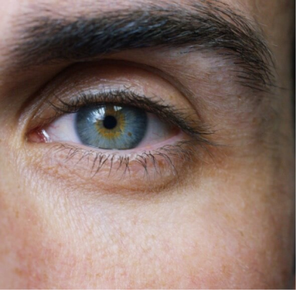
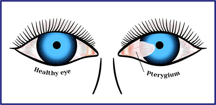
Pterygium (pronounced tur-IJ-ee-um), often called, “surfer’s eye” is an abnormal growth of vascular pink, fleshy tissue arising from the conjunctiva (a membrane that covers the white of the eye) towards the surface of the cornea. It commonly starts from the inner corner of the eye and grow towards the center of the eye. As it grows, it typically forms a triangle with the head of the pterygium towards the center.
[Pterygium By Jmvaras José Miguel Varas, MD (Own work) [GFDL (http://www.gnu.org/copyleft/fdl.html) or CC BY 3.0 (http://creativecommons.org/licenses/by/3.0)], via Wikimedia Commons]
Pterygium usually grows slowly throughout life and is a noncancerous lesion. They are benign (not malignant) tumors. It does not spread to other parts of the body. It may even stop growing after a certain point. In more advanced cases, though, it can cover the pupil of the eye and cause blurred and/or loss of vision.
Causes of Pterygium
The exact cause of Pterygium is still unknown. Many experts believe that, since it often occurs to people who live in warm climate areas, prolonged exposure to sunlight – particularly ultraviolet (UV) rays, increases the risk of pterygium. Pterygium is also more common to men than women, with fair or light-skinned complexion and those at who are at the age of 20-40 years old. It is also believed that chronic eye irritation and trauma from exposure to pollents, dust, sand, wind, and other environment stimuli can also add to the risk of pterygium formation.
Symptoms of Pterygium
In some cases, the pterygium has no symptoms other than its appearance – a raised pink, white, or red lesion on the eye that has blood vessels on the outer or inner edge of the cornea.
However, in most cases, symptoms of pterygium include itching, irritation, burning, tearing, gritty feeling or sensation of a foreign body in the eye. In the more advanced stage, symptoms of pterygium include redness, inflammation and decreased/blurred vision. When the lesion grows far enough onto the cornea (clear, outer layer of the eye), this may distort the shape of the cornea inducing astigmatism, as well as corneal scarring.
Pterygium Treatment
To date, there is no reliable medical treatment that can reduce or prevent pterygium progression. The only way to remove pterygium is via surgery.
Eyedrops
Pterygium usually doesn’t require treatment if symptoms are mild and doesn’t interfere with your vision. However, if irritation and redness causes discomfort, first line of treatment usually includes:
- lubricating eyedrops or ointments,
- vasoconstrictor eyedrops or,
- steriod eyedrops.
When the lesion progresses and causes persistent discomfort and interference wih vision or visual loss, Pterygium surgery can be done.
Pterygium Surgery
Pterygium surgery typically takes 30 to 45 minutes and is done as an outpatient procedure. Mild to moderate discomfort is expected after the procedure. Most people can resume activity within 2 days after the surgery process. More strenuous activities can be done usually after 2-4 weeks.
The first step to repair is excision of the pterygium, which can be done using different techniques, depending on the size and progression of the lesion. Conjunctival autograft or Amniotic membrane grafting are often done after excision to reduce complications and recurrence.
Conjunctival Autograft
Moving a part of the conjunctiva (mucous membrane that covers the front of the eye), and suturing the graft over the exposed scleral bed after excision of the pterygium.
Amniotic Membrane Grafting
Freeze-dried amniotic membrane (the surface membrane of the human placenta) is placed over the bare sclera, after excision of the pterygium.
Pterygium Surgery Risks
As with all other surgeries, Pterygium Surgery comes with some risks, including corneal scarring, perforation of the sclera, astigmatism, and the recurrence of a more aggressive lesion after removal.